Each clone is blotted in duplicate within each square. The 2304 small squares are subdivided into 16 units to which cDNA clones are blotted. ( C) Magnified image for one of the grid patterns shown in (B). These small squares are assigned a position by a numbered column and alphabetical row to identify positions of positive signals. ( B) A grid template overlay highlighting a portion of the 2304 squares in the GDA. ( A) A portion of one of six fields in the 22 × 22 cm GDA.

Only paired DNA targets (clones) with similar intensity signals, as determined by the imaging software, were considered positive.įigure 2 Representative gene expression pattern in human renal cortex probed with an array of 18,326 gene targets. The GenBank accession number, clone ID, and Human Gene Index number of full-length HTs for each expressed gene were retrieved electronically from Genome Systems. The identification of the expressed target gene was based on a distinct position and pattern in the array Figure 2d. A representative image of the high-density array gene expression is illustrated in Figure 2. Hybridization of the filter with each cDNA probe produces signals of variable intensity with each target cDNA clone. To determine the reproducibility of the hybridization profile, cDNA probes were prepared in duplicate from three renal specimens (probe 6a, 6b 7a, 7b and 9a, 9b) and hybridized to two separate filters. To establish comprehensive gene expression profile of the renal cortex, the dCTP-labeled cDNA probe from each renal specimen was hybridized to a high-density cDNA array, with 18,326 paired targets.
#HYBAID MINI HYBRIDIZATION OVEN MANUAL SOFTWARE#
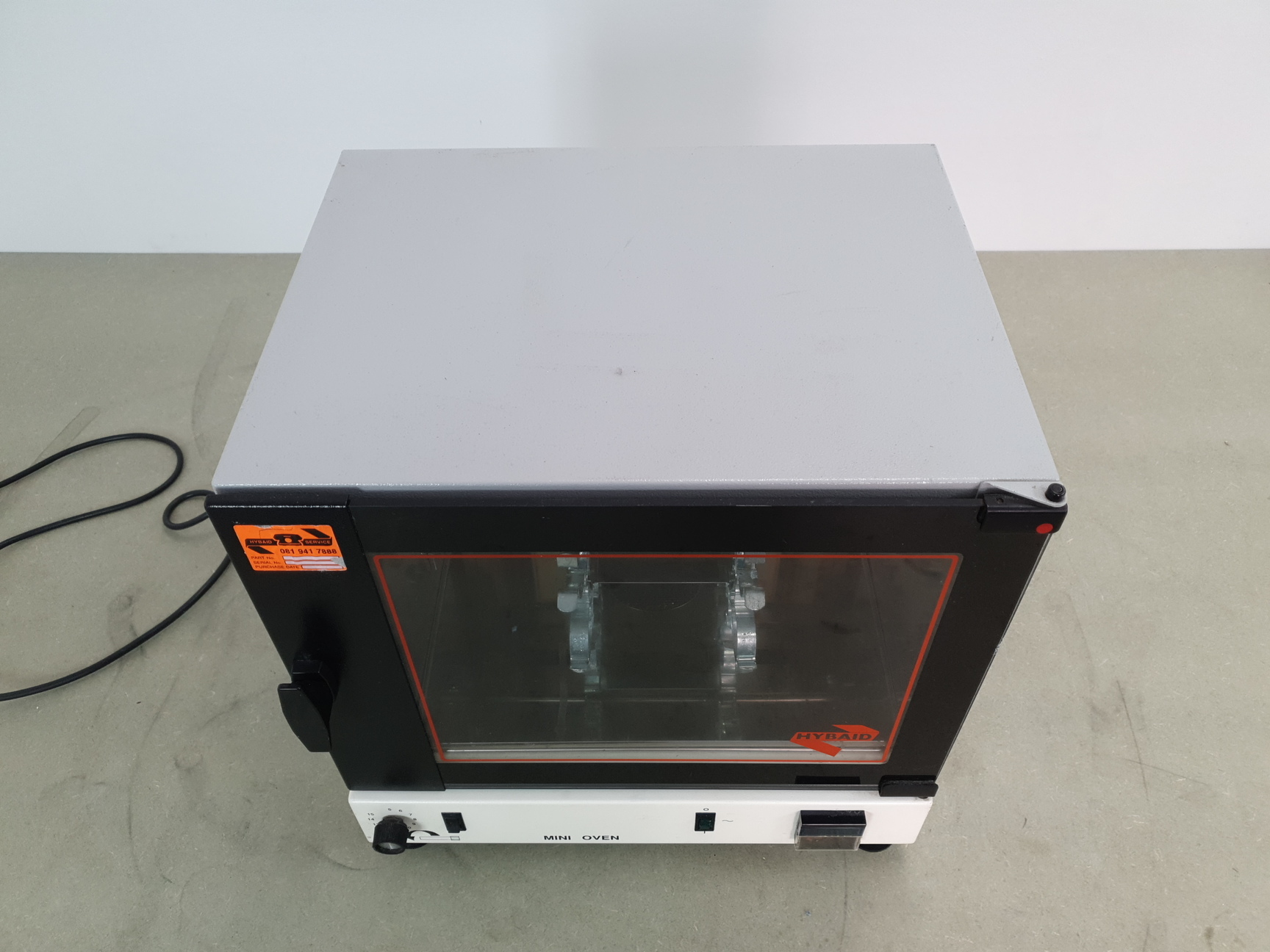
Signals developed on the imaging plates were processed for quantitative analysis using imaging software specialized for high-density array analysis (Array Vision™ IMAGING Research Inc., St. The plastic-wrapped membrane was exposed to a BAS-MS2040 imaging plate (Fuji Photo Film Co., Ltd., Kanagawa, Japan) for 48 to 72 hours, and then the image was read by a BAS-2500 phosphorimager (Fuji Photo Film). All the washes were performed in the hybridization bottles at 17 r.p.m. After hybridization, the membrane was washed twice for 10 minutes at room temperature with 2 × saline-sodium phosphate-EDTA (SSPE) buffer supplemented with 0.1% SDS, followed by extensive wash with 1 × SSPE and 0.1 × SSPE buffer for 20 minutes at 65☌ twice and once, respectively. The solution was added to the membrane prehybridized in the same buffer and was incubated at 42☌ for 16 hours with continuous rotation (10 r.p.m.) in a hybridization oven (HYBAID). The radiolabeled cDNA probe (20 × 10 6 cpm) was added to 10 mL of hybridization buffer.

Louis, MO, USA), was used for the hybridization studies. The remainder of the probe solution was used immediately for the hybridization.Ī commercial array of high-density cDNA clones, Gene Discovery Array version 1.3 GDA-Filter 1 (Genome System Inc., St.

A small aliquot from each probe was stored at -80☌ before analysis by gel electrophoresis. The cDNA was labeled with 25 μCi dCTP (NEN Life Science Products, Boston, MA, USA) using the Prime-It® II Random Primer Labeling Kit (Stratagene, La Jolla, CA, USA), followed by purification by CHROMA SPIN-100 Column (Clontech Laboratories, Inc., Palo Alto, CA, USA). Complex cDNA probe was prepared from poly A + mRNA purified from 25 μg of total RNA by Dynabeads oligo (dT) 25 (Dynal, Lake Success, NY, USA) using SuperScript RNase H - reverse transcriptase (Life Technology, Gaithersburg, MD, USA). Total RNA was extracted from homogenate of the renal cortex using TRIzol™ Reagent (GIBCO BRL, Gaithersburg, MD, USA) and was then treated with DNase I (Promega Corp., Madison, WI, USA) to eliminate potential contamination with genomic DNA. From a collection of 30 nephrectomies, we selected nine renal tissue samples that showed no pathological features by microscopic examination Table 1.

An uninvolved cortical section of a nephrectomy from patients who underwent renal resection was snap frozen in liquid nitrogen and stored at -80☌.


 0 kommentar(er)
